
What is photobleaching, and how does it impact medical imaging?
Photobleaching poses a substantial challenge in medical imaging, potentially compromising the accuracy and reliability of fluorescent-based techniques of various applications.
It’s important to understand how photobleaching works before taking steps to protect fluorescent signals.

Image: fluorescence microscopy
That way, medical applications can enhance the quality of their imaging-based analyses and ensure more accurate diagnostic and research outcomes.
In this blog, you’ll learn about the ins and outs of photobleaching, its causes and implications, and how you can overcome this major challenge.
What is photobleaching?
Photobleaching happens when fluorophores, the molecules responsible for fluorescence, are exposed to continuous or intense light. Photochemical alterations cause permanent changes to the molecule, making it lose its ability to fluoresce.
This degradation process is a large concern in fluorescence microscopy and related imaging methodologies, where the accuracy of fluorescent signals is critical.
Implications of photobleaching in medical imaging
Now that we understand what photobleaching is – let’s quickly go through its implications. The reduction of the fluorescent signal due to photobleaching poses serious challenges for medical imaging. This is especially true in diagnostics or research, where detecting fluorescent signals is important for identifying specific cells, structures, or molecules, leading to skewed data.
Let’s look at some of the other medical imaging issues caused by photobleaching.
![]()
Image: a close-up view of light-induced damage in biological samples
Reduced diagnostic accuracy
Photobleaching leads to the fading of fluorescent signals, which are crucial for identifying and analysing specific cellular components or markers in medical imaging. The reduction in signal intensity can lead to false negatives, where the presence of a target molecule or cell is undetected. It may trigger incorrect diagnoses or the oversight of critical information.
Compromised longitudinal studies
Photobleaching can hinder the ability to accurately track changes while monitoring the progression of a disease or the response to a treatment. The weakening of fluorescent signals complicates the image comparison across time points, affecting the reliability of longitudinal analyses.
To mitigate this, different fluorophores can be used for longitudinal studies. However, this isn’t always straightforward or ideal. Switching fluorophores can introduce variability in the imaging process, as different fluorophores may have different photophysical properties, leading to inconsistencies in the data.
Potential cost and time increase
To address the loss of signal from photobleaching, additional fluorophores may be required, or imaging sessions might need to be repeated. This could result in higher costs for reagents and consumables, along with more time spent on imaging and analysis. However, whether these additional costs are considered a drawback depends on the specific scenario. For instance, it may be justified in some as they contribute to more reliable data. In other settings, it may lead to a considerable cost burden.
Altered sample integrity
The methods used to counteract photobleaching, such as increasing the concentration of fluorophores or using more intense illumination, can have detrimental effects on the biological samples being studied. High-intensity light can cause phototoxicity, affecting the viability of cells and potentially inducing artificial changes in their behaviour or properties, which could skew research findings or diagnostic outcomes.
Limitations on quantitative analysis
Photobleaching affects the ability to perform quantitative fluorescence imaging, a technique for accurately measuring the concentration of specific molecules within a sample. The loss of signal intensity over time can lead to an underestimation of target molecule concentrations. It can also impact the quantitative accuracy of the analysis, resulting in inaccurate conclusions about the composition of the sample.
How to reduce photobleaching in medical imaging
Limiting illumination to the focal plane
Reducing the overall exposure of non-target areas becomes possible by confining the illumination and excitation of fluorophores strictly to the area of interest. It ensures that only the regions requiring analysis are illuminated, minimising unnecessary light exposure to surrounding areas.
It also conserves the fluorophores’ fluorescence capabilities while enhancing the clarity and contrast of the acquired images.
Lowering peak intensity
Adjusting the intensity of illumination to the lowest effective level decreases the energy absorbed by fluorophores, reducing the likelihood of covalent modifications that lead to photobleaching. It requires careful measurement to make sure that the illumination is enough to excite the fluorophores without inducing extreme stress that could trigger photobleaching.
Moreover, this contributes to preserving the structural integrity of the biological specimens under examination, preventing damage that could result from high-intensity illumination.
Pulsed illumination
Implementing pulsed rather than continuous illumination provides intervals during which fluorophores can recover, diminishing the cumulative impact of light exposure. It involves intermittently applying light to the specimen, allowing periods of darkness between pulses. These breaks in illumination help reduce the overall energy absorbed by the fluorophores, extending their functional lifespan.
It also maintains the fluorescent signal’s intensity over longer periods. Pulsed illumination can be of great help in live-cell imaging, where prolonged exposure to continuous light gives rise to photobleaching risks.
Key camera features that help mitigate photobleaching
Size of the aperture
A larger aperture in cameras ensures that more light enters the system. It reduces the need for longer exposure times, which can otherwise increase the risk of photobleaching in fluorescent imaging.
Transmission components (mirrors and filters)
High-quality mirrors and filters ensure proper transmission of light to the sensor. These components minimise light loss by optimisng the light path. It minimises the intensity and duration of illumination required, which, in turn, mitigates photobleaching.
Global shutter and trigger
Cameras with a global shutter capture the entire image at once, avoiding the distortion or motion blur that might require repeated imaging. The setup can be coupled with trigger mechanisms that synchronise image capture with specific actions. This minimises unnecessary exposure to light and reduces the possibility of photobleaching.
Monochrome sensor
Monochrome sensors are more sensitive to light than colour sensors, especially in detecting fluorescence. They require less illumination for the same signal strength, which helps preserve fluorophores and prevent premature photobleaching.
Quantum Efficiency (QE)
Cameras with higher QE convert more of the incoming light into electrical signals. This ensures accurate imaging with lower light intensity, mitigating the photobleaching risks.
Signal-to-Noise Ratio (SNR)
A higher SNR means that the camera can provide image clarity at lower exposure and illumination levels, which helps minimise photobleaching.
Product Spotlight
STDRIVEG611Q
STMicroelectronics

| SKU: | 497-STDRIVEG611QTR-ND |
|---|---|
| Stock: | 0 |
| Cost: | $2.63 |
TBF10SL-4PS-B
ITT Interconnect Solutions
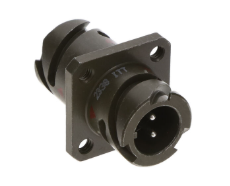
| SKU: | 1003-TBF10SL-4PS-B-ND |
|---|---|
| Stock: | 50 |
| Cost: | $45.59 |
CAA572C0G3A663J640LJ
TDK Corporation
| SKU: | |
|---|---|
| Stock: | 1037 |
| Cost: | $9.16 |
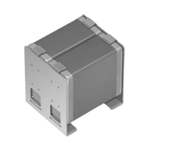
R30-3002002
Harwin

| SKU: | 952-1510-ND |
|---|---|
| Stock: | 6545 |
| Cost: | $0.76 |
NRF54L15-QFAA-R
Nordic Semiconductor
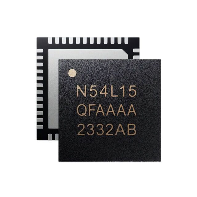
| SKU: | 4823-NRF54L15-QFAA-RTR-ND |
|---|---|
| Stock: | 0 |
| Cost: | $2.39 |
STDRIVEG611Q
STMicroelectronics
| SKU: | 497-STDRIVEG611QTR-ND |
|---|---|
| Stock: | 0 |
| Cost: | $2.63 |
TBF10SL-4PS-B
ITT Interconnect Solutions
| SKU: | 1003-TBF10SL-4PS-B-ND |
|---|---|
| Stock: | 50 |
| Cost: | $45.59 |
CAA572C0G3A663J640LJ
TDK Corporation
| SKU: | |
|---|---|
| Stock: | 1037 |
| Cost: | $9.16 |
R30-3002002
Harwin
| SKU: | 952-1510-ND |
|---|---|
| Stock: | 6545 |
| Cost: | $0.76 |
NRF54L15-QFAA-R
Nordic Semiconductor
| SKU: | 4823-NRF54L15-QFAA-RTR-ND |
|---|---|
| Stock: | 0 |
| Cost: | $2.39 |
STDRIVEG611Q
STMicroelectronics
| SKU: | 497-STDRIVEG611QTR-ND |
|---|---|
| Stock: | 0 |
| Cost: | $2.63 |